About the Practical Course
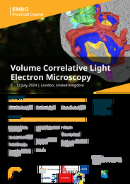
In 2023, volume Electron Microscopy (vEM) was named as one of the seven technologies to watch by Nature. vEM is a term that describes a group of techniques that reveal the 3D ultrastructure of cells and tissues through continuous depths greater than 1 micron. vEM techniques require correlative workflows using light and/ or X-ray microscopy to target the limited fields of view that can be imaged by EM. These workflows are known as volume Correlative Light and Electron Microscopy (vCLEM). This practical course on vCLEM is an evolution of the previous EMBO CLEM courses, taking into account the huge progress in the field in a very short space of time and with an updated programme that reflects the current state of the art in correlative and multimodal imaging. A critical aspect of the course will be the focus on software for image acquisition, processing and analysis, and FAIR data sharing through public image archives. Four workflows will be covered on the course:
Workflow 1: Correlative Array Tomography
Tissue containing fluorescent protein tags will be sliced using a vibratome, imaged using a Laser Scanning Confocal Microscope with enhanced resolution, embedded using the Ellisman/ Deerinck protocol, sectioned using an ultramicrotome with manual and semi-automated section retrieval, and imaged in the SEM with AT acquisition software.
Workflow 2: X-ray targeting for Block Face Imaging
Heavy metal-stained, resin-embedded tissue will be imaged by microCT to locate a region of interest, trimmed to the plane of interest using Crosshair software and a motorised ultramicrotome, and the ROI imaged using Serial Block Face Scanning Electron Microscopy with SBEMimage open-source image acquisition software.
Workflow 3: High pressure freezing to electron tomography and FIB-SEM
Cells expressing fluorescent proteins will be imaged live on sapphire discs in a Laser Scanning Confocal Microscope and vitrified using a high pressure freezer with fast transfer capabilities. Samples will be freeze substituted, sectioned and imaged by TEM tomography using a transmission electron microscope with serial EM acquisition software. In parallel, FS protocols that incorporate heavy metals for vEM workflows and FIB-SEM image acquisition will be discussed.
Workflow 4: Visual Proteomics CLEM Kit
A monolayer of cells labelled with fluorescent proteins, tracker dyes and/or Alexa dyes will be prepared using a non-HPF In-Resin Fluorescence (IRF protocol). The resulting resin blocks will be serial sectioned onto substrates, imaged using serial super resolution light microscopy, followed by array tomography in a scanning electron microscope, to produce high resolution volume CLEM overlays. We will also show an alternative workflow to target the florescent regions, where the block is imaged with a confocal microscope and the fluorescence 3D map is used to target a specific cell of interest for vEM imaging.
Workflow 5: Software
Experts in open-source software and public image archiving will train participants in how to: Understand and use AI methods for segmentation, including the use of manual annotation and citizen science to gather training data; align LM to EM data using CLEM-Reg software; handle multimodal image data in MOBIE; and deposit correlative image data into EMPIAR and the BioImage Archive.
About EMBO Courses and Workshops
EMBO Courses and Workshops are selected for their excellent scientific quality and timelines, provision of good networking activities for all participants and speaker gender diversity (at least 40% of speakers must be from the underrepresented gender).
Organisers are encouraged to implement measures to make the meeting environmentally more sustainable.


