
About the Practical Course
Cryo electron microscopy (cryo EM) is a major structural biology method for studying macromolecular complexes and cellular structures in their native states. It can be used to sort out heterogeneous conformations to capture steps in the operation of macromolecular machines. Stable high resolution cryo microscopes, direct electron detectors and automated systems for data collection now allow routine biomolecular structure determination at near-atomic resolution. In structural studies by electron tomography the resolution has improved to the subnanometer range. Fitting of individual structures into cryo EM maps of macromolecular complexes reveals interactions of their components and conformations of different functional states. Hybrid approaches combining EM with optical microscopy and dynamic studies uncover biological mechanisms. New approaches in analysis of thin cells and cryo-lamellae by tomography extend the range of EM from isolated complexes to cellular structures in situ.
The aim of this EMBO Practical Course is to teach the basic principles and practical aspects of image processing to structural and molecular biologists wishing to determine macromolecular structures by cryo EM. The course will concentrate on processing of high-resolution image data, for both single particle images and tomograms, and will be aimed at advanced PhD students and postdocs using cryo EM for structural analysis.
Image credits: Katerina Toropova and Anthony Roberts (header)
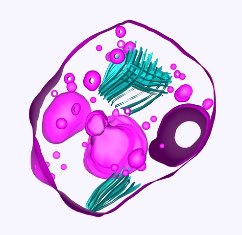
Ya Zhou and Helen Saibil (small figure)
About EMBO Courses and Workshops
EMBO Courses and Workshops are selected for their excellent scientific quality and timelines, provision of good networking activities for all participants and speaker gender diversity (at least 40% of speakers must be from the underrepresented gender).
Organisers are encouraged to implement measures to make the meeting environmentally more sustainable.


.jpeg)
.jpg)